OD5
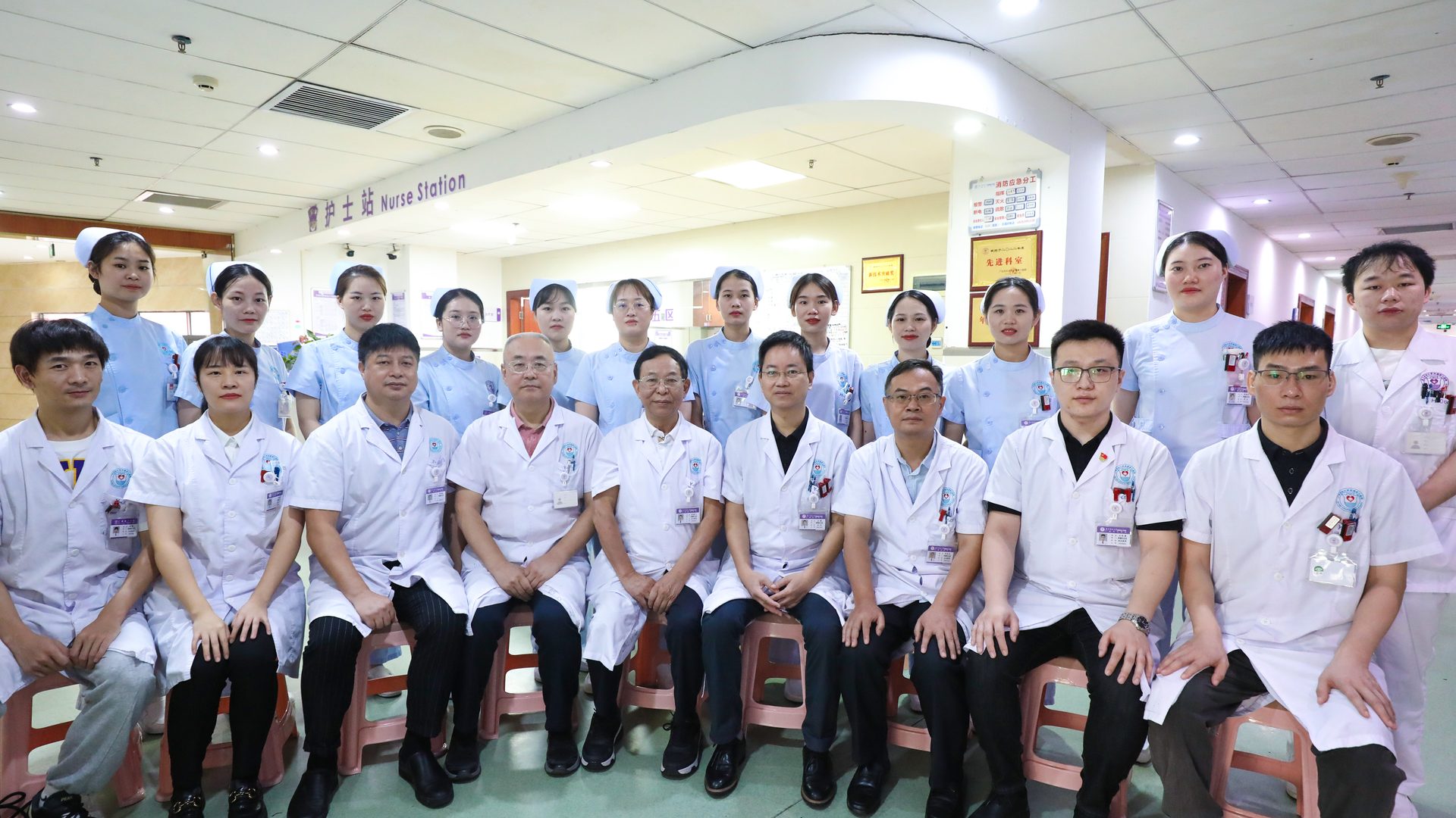
Oncology Ward 5Guangzhou Fosun Chancheng Hospital of Guangdong Pharmaceutical University (Guangzhou Xinshi Hospital)
WhatsApp +852 4743 1548
Oncology Ward 5 · International Center for Minimally Invasive Oncology

Comprehensive Minimally Invasive Care for Malignant Tumors
Centered on the Luo Pengfei Programmed Oncology Approach

Led by Professor Luo Pengfei, a renowned interventional oncology specialist, our interventional research team provides individualized, standardized and specialized comprehensive care for patients with a wide range of advanced solid tumors.

WhatsApp +852 4743 1548

Submit your imaging studies and medical history — our specialist team will assess which minimally invasive options may be appropriate

50bedsLicensed inpatient beds
30+expertsOncology specialist panel
9deptsIn-hospital collaborator departments
1500sqmInternational medical center floor area
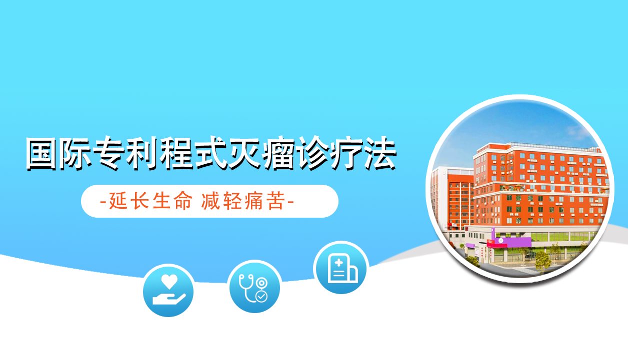
International Center for Minimally Invasive Oncology Diagnosis and Treatment
Department Overview

A Key Minimally Invasive Oncology Discipline of the Hospital

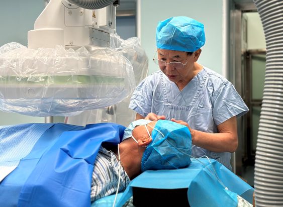
Oncology Department V is a key discipline for minimally invasive oncology at the hospital, built around the Luo Pengfei Programmed Oncology interventional research team. The team's three chief physicians are Prof. Luo Pengfei (President, Southern Institute of Interventional Radiology), Prof. Wang Jian (Department Director) and Prof. Cui Zhe. In September 2010, renowned oncology expert Prof. Luo Pengfei received the "Outstanding Contribution Award" at the 9th National Interventional Radiology Academic Conference. The department's mission: individualized, programmed and specialized comprehensive treatment for malignant tumors — to control the disease, improve quality of life, care for life, and serve with empathy. Patients with intermediate to advanced solid malignant tumors are suitable for our department, including liver, lung, gastric, breast, colorectal and esophageal cancers, malignant lymphoma, malignant melanoma, urological, gynecological, head & neck, intracranial and bone tumors, soft-tissue and retroperitoneal tumors, metastatic cancer, benign uterine fibroids and others.

A Key Minimally Invasive Oncology Discipline of the Hospital
◆

Team Leadership

Care is directed by an interventional research team of professors including Luo Pengfei, Wang Jian and Cui Zhe, with the chief physician performing the procedures personally.

◆

Multi-Technology Integration

A combined use of minimally invasive techniques including programmed multi-level drug-eluting microspheres, radioactive seed implantation, argon-helium knife cryoablation, stent implantation and non-invasive deep tumor hyperthermia.

◆

Derived from a Patented Therapy

Each technique has been developed from the Luo Pengfei Programmed Oncology Approach, a therapy system holding international patents in the field of interventional oncology.

◆

Broad Applicability

Our scope of care covers liver, lung, gastric, breast, colorectal and esophageal cancer, malignant lymphoma, malignant melanoma, and tumors of the urinary system, gynecological system, head and neck, intracranial region, bone, soft tissue and retroperitoneum, as well as metastatic cancer and certain benign tumors.

Specialist Team

Leaders in Minimally Invasive Tumor Treatment

An interventional research team led by three chief physicians, providing multidisciplinary team (MDT) support for complex cases.

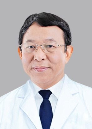
Luo Pengfei

Luo Pengfei

Chief Physician / Professor
Founder of the Luo Pengfei Programmed Oncology Approach
  • Deputy Director, Guangdong Cancer Prevention and Treatment Center
  • Director, Imaging & Interventional Center, Guangdong Provincial People's Hospital · Lifetime Professor
  • Recipient of the State Council Expert Contribution Allowance
Over 40 years of clinical experience in solid tumors including liver, lung, pancreatic, cholangiocarcinoma, head & neck, bone metastases and colorectal liver metastases.
Received the Outstanding Contribution Award at the 9th National Interventional Radiology Conference (2010); international patent LU501835.
Wang Jian

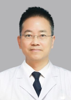
Wang Jian

Chief Physician
Inheritor of the Programmed Micro-needle Technique · Team Lead
  • Chairman and Lead Operator, Luo Pengfei & Wang Jian Interventional Research Team
  • Standing Director, China Women's & Children's Interventional Alliance
  • Committee Member, Hepatocellular Carcinoma Committee, Guangdong Anti-Cancer Association
Integrates superselective arterial chemoembolization, radioactive seed implantation, physical ablation and vascular / esophageal / biliary stenting. Over 30 years in interventional practice.
Recipient of Science & Technology Progress Awards from Foshan and Shunde municipal governments.
Cui Zhe

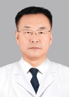
Cui Zhe

Chief Physician, Medical Oncology
Lingnan Distinguished Physician · Research Team Board Member
  • Recipient of the Lingnan Distinguished Physician title
  • Board Member, Luo Pengfei Programmed Oncology Research Team
  • Focused on minimally invasive interventional therapy and systemic oncologic care
Specialises in early diagnosis and minimally invasive treatment of benign and malignant tumours, alongside chemotherapy, targeted therapy, immunotherapy and multidisciplinary care.
Published in leading radiology and interventional journals; recipient of provincial medical science progress awards.
Treatment Techniques

Five Advanced Techniques of the Department

A single or combined minimally invasive approach is selected based on lesion location, stage and the patient's general condition.

01

Luo Pengfei Programmed Multi-Level Drug-Eluting Microsphere Technique (Patented)

An innovative patented technique enabling precise drug loading, with comprehensive drug-eluting embolization from the distal peripheral vessels to the proximal main trunk.

02

Radioactive Seed Implantation

Also known as internal radiation therapy (brachytherapy). Radioactive seeds are placed into the lesion under CT guidance to act continuously within the tumor.

03

Cryoablation

Including argon-helium knife cryoablation and microwave thermal ablation, using ultra-low or ultra-high temperatures to induce necrosis of tumor tissue.

04

Stent Implantation

Used to relieve obstruction and stenosis with minimal trauma and faster recovery, helping improve feeding, jaundice and blood supply.

05

Non-Invasive Deep Tumor Hyperthermia

Non-invasive external radiofrequency heating; deep heating assists in devitalizing tumor cells.

Core Technology in Detail

Luo Pengfei Programmed Multi-Level Drug-Eluting Microsphere Technique (Patented)

Internationally Patented Technology

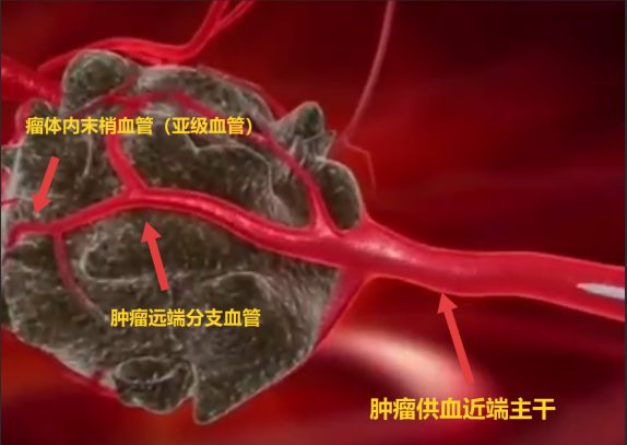
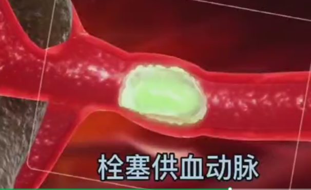
The interstitial matrix of a tumor is denser than that of normal tissue, forming a physical barrier. In conventional treatment, drugs often fail to penetrate or penetrate insufficiently deep, so tumor cells in the deeper layers are not exposed to an adequate drug concentration. Under superselective catheterization, the Luo Pengfei programmed multi-level technique delivers a high concentration of drug into the tumor and embolizes the tumor's sub-level vessels. The drug is retained within the tumor, creating a local concentration gradient that uses physical pressure to drive the agent deeper into the interstitium, thereby reaching tumor cells further inside the lesion.

Conventional intervention: drug loading at the proximal main tumor-feeding trunk
Luo Pengfei programmed technique: comprehensive drug-eluting embolization from the peripheral vessels to the proximal main trunk

Foundation in an Internationally Patented Therapy

Derived from the Luo Pengfei Programmed Oncology Approach. This therapy follows a step-by-step rationale of tumor control, tumor shrinkage and tumor devitalization, aiming to inactivate tumor biological activity and interrupt pathways of recurrence and metastasis.

Levels of Vascular Intervention

Covers three levels: the proximal main tumor-feeding trunk, the distal branch vessels, and the peripheral vessels within the tumor itself.

Graded Drug-Eluting Embolization

Superselective catheterization can be performed down to fourth-level arteries and their sub-branches, enabling precise embolization of third-level, fourth-level and sub-level vessels.

Advantages of Superselective Technique

Helps identify and address uncommon or potential small collateral tumor-feeding arteries in a timely manner.

Drug Retention Effect

After loading, the high-concentration drug is retained within the tumor and continues to act; the PPT source material records a duration of approximately 7–14 days.

Safeguards in Delivery

Procedures are performed personally by the chief physician.

Radioactive Seed Implantation
Technology in Detail

Radioactive Seed Implantation

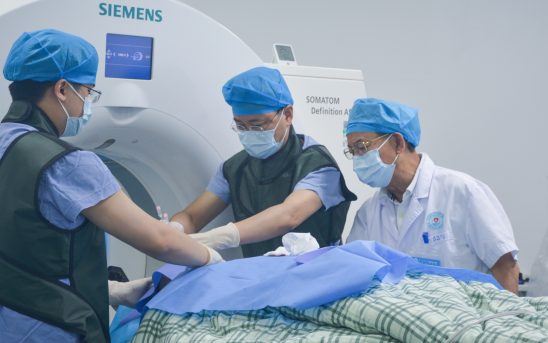
Also known as internal radiation therapy (brachytherapy). Under CT guidance, a fine needle is advanced precisely into the tumor, and radioactive Iodine-125 seeds are delivered through the needle so that they are finely distributed throughout the lesion. The spontaneous, continuous radiation emitted by the seeds acts on the tumor cells.

  • Precise, image-guided puncture for seed placement
  • Seeds release radiation continuously within the lesion
  • Suitable for certain solid tumors in difficult locations that are not amenable to physical ablation
  • Clinically used in lung cancer, liver cancer, brain tumors, breast cancer, pancreatic cancer, head and neck cancer, esophageal cancer, mediastinal tumors, bone tumors, cervical cancer and others
Cryoablation
Technology in Detail

Cryoablation

An ablation probe is inserted into the tumor under CT guidance, using ultra-high or ultra-low temperatures to cause protein denaturation and necrosis of tumor cells, achieving the treatment purpose.

  • Microwave thermal ablation
  • Endobronchial radiofrequency ablation
  • Argon-helium knife cryoablation

Applicable to solid tumors such as lung, liver and breast cancer, with minimal trauma.

Stent Implantation
Technology in Detail

Stent Implantation

For stenosis and obstruction of passages caused by tumors, stent implantation restores patency and helps improve the patient's quality of life.

  • Malignant Biliary Obstruction:A biliary stent is implanted via percutaneous transhepatic puncture — minimally invasive, and helpful in relieving jaundice and supporting recovery of liver function.
  • Esophageal Stenosis and Obstruction:Esophageal stent implantation may ease tumor-related esophageal stricture and obstruction, helping restore the ability to eat.
  • Vascular Stenosis and Occlusion:Vascular stent implantation can restore blood flow and improve organ perfusion.
Non-Invasive Deep Tumor Hyperthermia
Technology in Detail

Non-Invasive Deep Tumor Hyperthermia

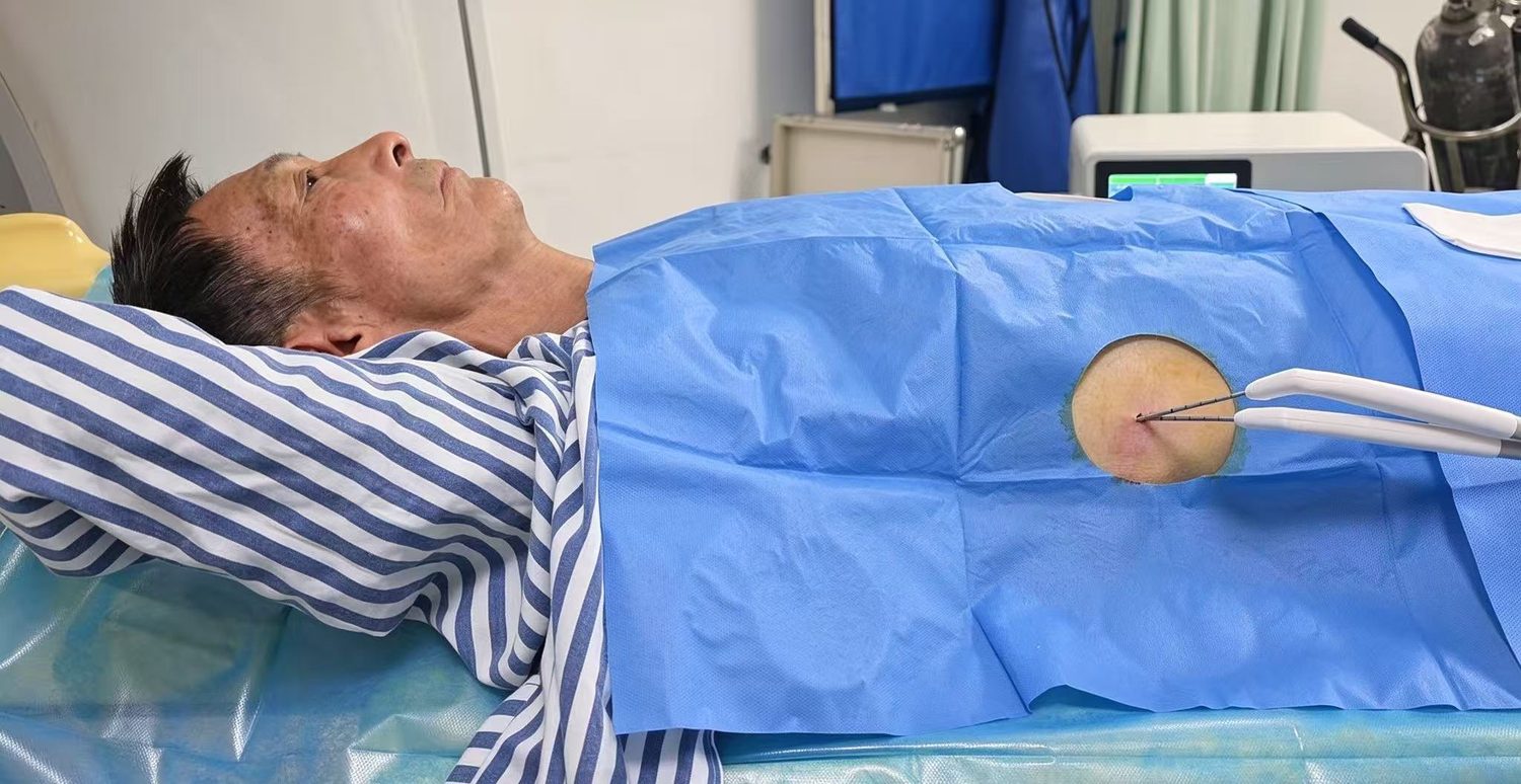
Using an external radiofrequency hyperthermia unit, deep-seated lesions are heated in a non-invasive manner. It is an important adjunct within the minimally invasive treatment system.

  • Advantages of Non-Invasive Hyperthermia:No incision and no ionizing radiation; deep heating acts on cancer cells.
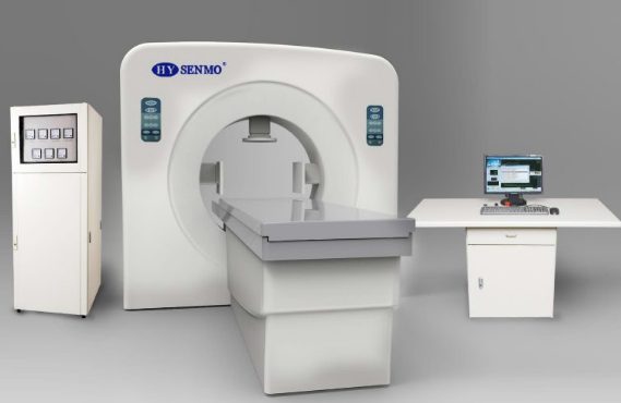
  • Equipment and Heating Method:The HY7000-I radiofrequency hyperthermia unit uses four time-shared rotating applicators, supported by a protocol for fat heat accumulation and skin surface protection.
  • Treatment Parameters:Target heating above 39°C per session, sustained for approximately 40 minutes; a recommended course consists of 6 sessions.
  • Scope of Application:Primary, recurrent, deep-seated or superficial solid tumors and metastatic tumors, excluding those of the head and neck.
Clinical Records

The Clinical Course of Selected Complex Cases

The following are records of international patients treated by the department. They are provided only to illustrate the clinical process and methods; individual circumstances differ and nothing here constitutes any guarantee of treatment outcome.

Bladder cancer · recurrent gross hematuria
Ms. L, 72 years old, Indonesian
Bladder cancer · recurrent gross hematuria
On admission
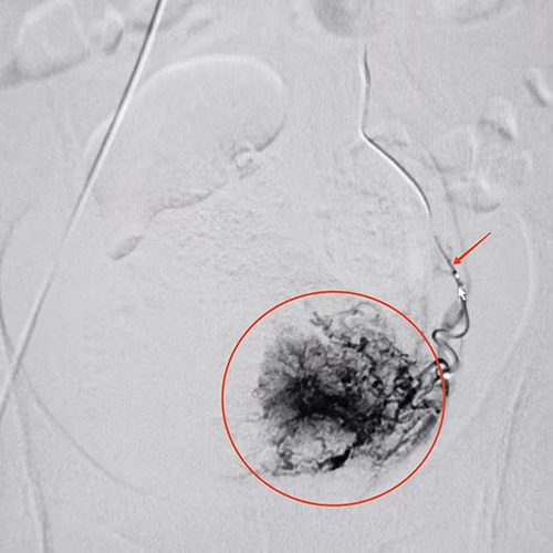
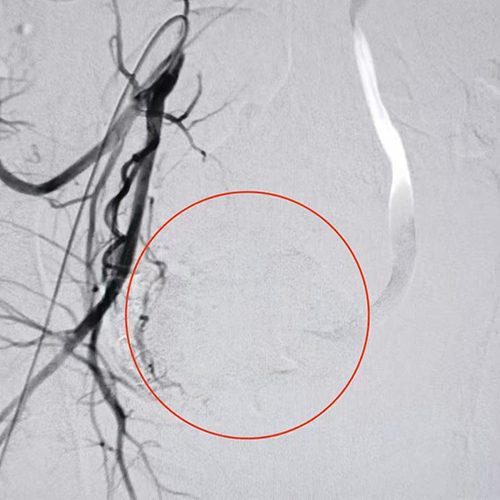
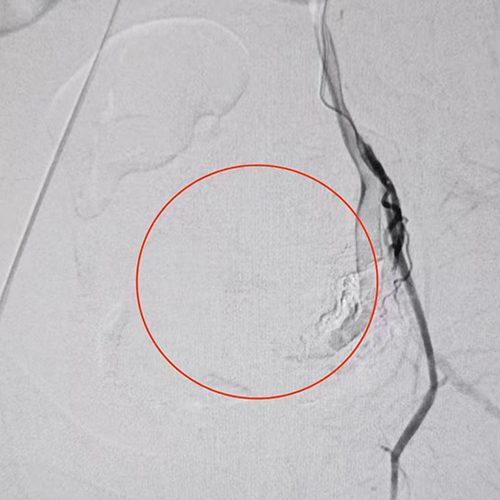
She had recurrent gross hematuria for over a year beginning in 2024. She was diagnosed with bladder cancer and received approximately six months of comprehensive treatment. After hematuria worsened over the past month, she was transferred to our department. On admission, she had total-stream hematuria and severe anemia (hemoglobin 56 g/L). After red blood cell transfusion, emergency TACE was performed to control bleeding while also addressing the tumor.
Imaging & interventional records
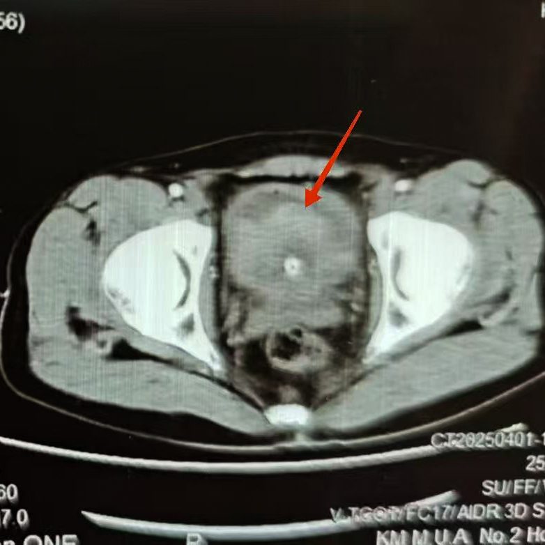
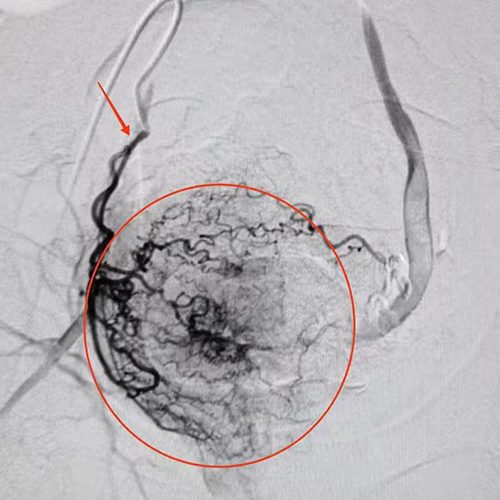
Figure 1: Pre-procedure CT demonstrating the bladder tumor. Figures 2–3: Bilateral vesical artery angiography showing extensive tumor staining and signs of contrast extravasation. Figures 4–5: Post-embolization angiography following bilateral vesical artery infusion embolization, with disappearance of tumor staining.
Post-procedure observation
Hematuria reduced rapidly after the procedure, with urine clearing by the next day's round.
Technical notes
For conditions such as bladder cancer bleeding, the Luo Pengfei programmed multi-level drug-eluting technique, under superselective catheterization, helps identify uncommon or potential small collateral feeding arteries at the earliest opportunity, enabling drug delivery targeted at sub-level vessels.
Tongue cancer (squamous cell carcinoma)
Ms. Z, 54 years old, Malaysian
Tongue cancer (squamous cell carcinoma)
On admission
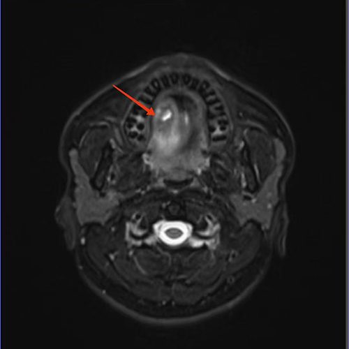
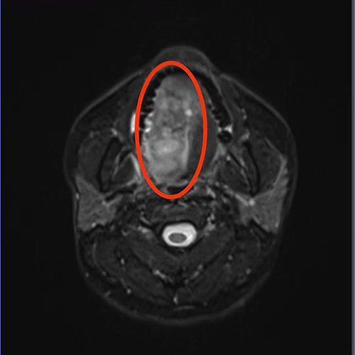
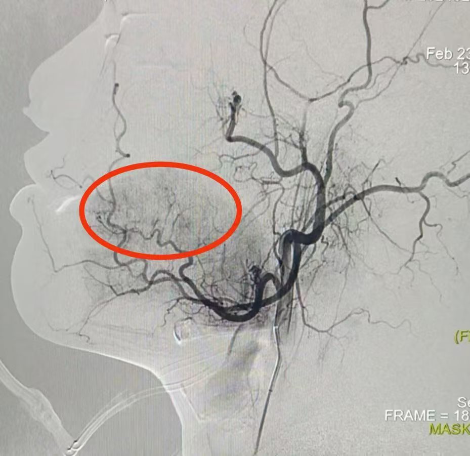
She noticed a white patch on the right side of the tongue one year ago, which gradually developed into a mass that progressively enlarged with tenderness over six months. After admission, a needle biopsy revealed squamous cell carcinoma. Examination showed a space-occupying lesion of the right tongue crossing the midline, and right external carotid artery angiography demonstrated tumor staining in the tongue.
Imaging & interventional records
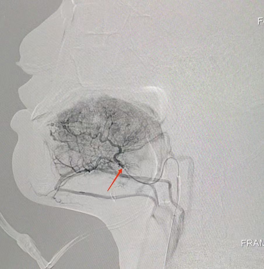
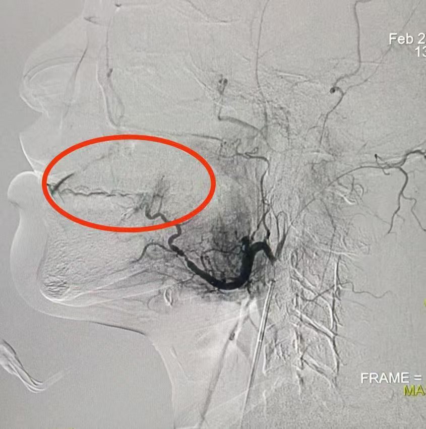
Figures 1–2: MR showing the right tongue space-occupying lesion crossing the midline. Figure 3: Right external carotid angiography showing tumor staining. Figure 4: Superselective lingual artery angiography showing abundant tumor vessels and staining. Figure 5: Post-embolization angiography with disappearance of tumor staining.
Post-procedure observation
Infusion embolization was completed, and tumor staining disappeared.
Technical notes
Under superselective catheterization, the multi-level drug-eluting technique delivers a high drug concentration into the tumor and embolizes sub-level vessels. The drug retained within the tumor forms a local concentration gradient that uses physical pressure to drive penetration into the deeper interstitium.
Right lung squamous cell carcinoma with multiple metastases
Mr. C, 68 years old, Singaporean
Right lung squamous cell carcinoma with multiple metastases
On admission
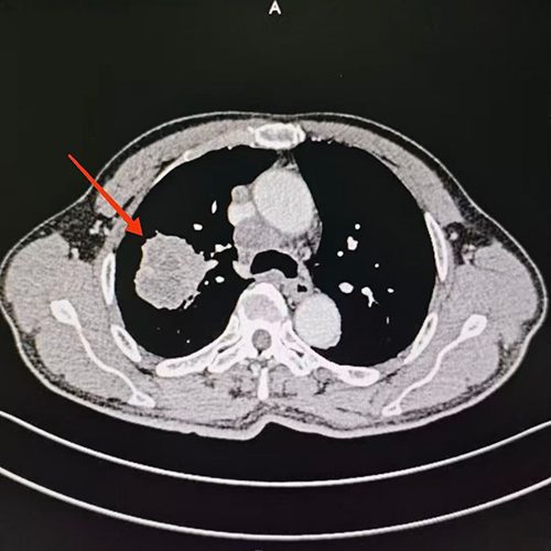
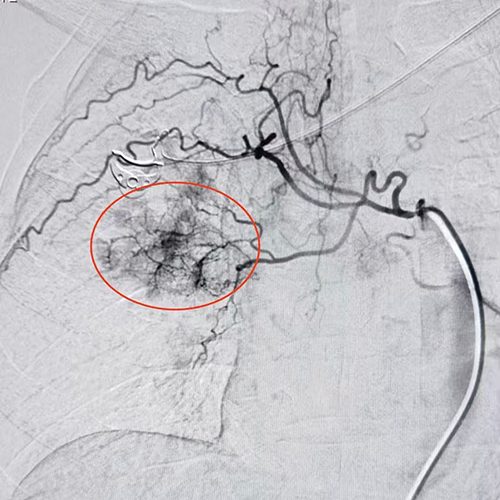
He was diagnosed one month ago with right lung squamous cell carcinoma with multiple metastases. After extensive research, he returned to China to seek care at our hospital. The team developed separate interventional plans for the primary lesion and the metastatic lesions.
Imaging & interventional records
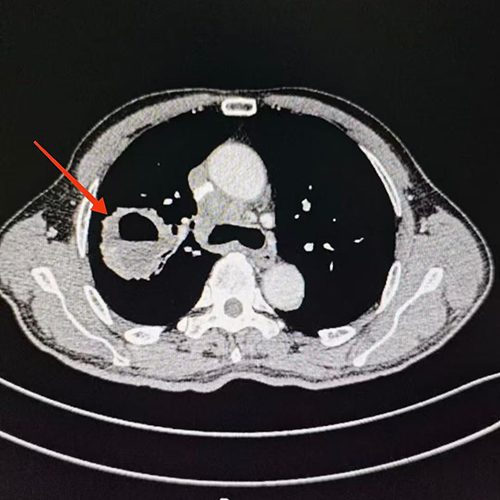
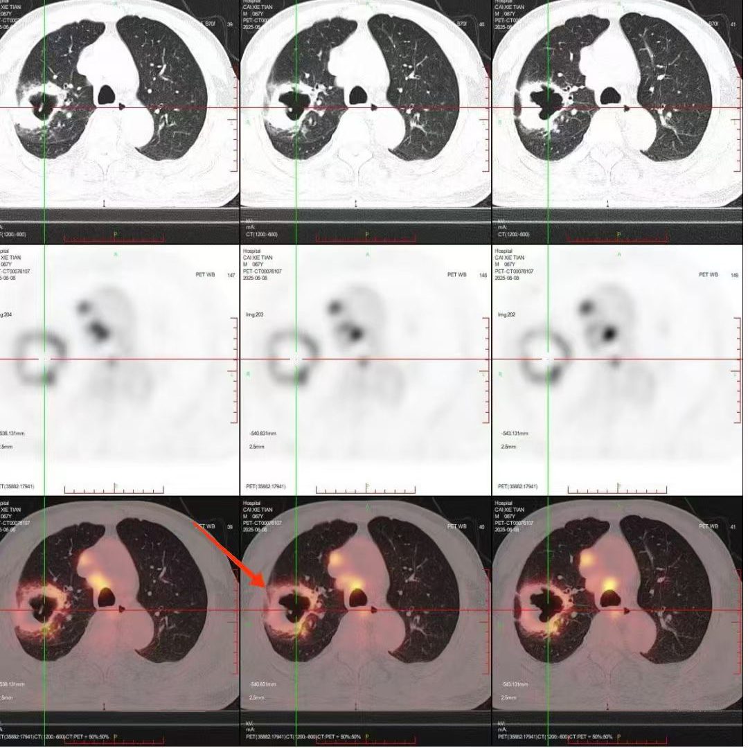
Figures 1–2: Initial CT demonstrating the right lung tumor and the first bronchial artery angiography. Figures 3–5: Follow-up showing tumor necrosis with cavity formation, and bronchial artery angiography showing reduced tumor vessels and staining.
Post-procedure observation
Follow-up showed tumor necrosis with cavity formation, and reduced tumor vessels and staining.
Technical notes
The more peripheral the vascular level, the finer the branches. Conventional interventional technique requires selective catheterization to third-level branch arteries, whereas the programmed multi-level drug-eluting technique requires superselective catheterization to fourth-level arteries and their sub-branches, which favors higher intratumoral drug concentration and greater embolization extent.
International Medical Center

Deepening Oncology Care, Opening a New Chapter in International Medicine

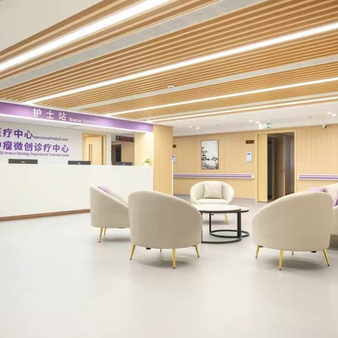
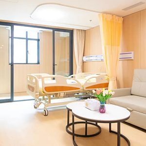
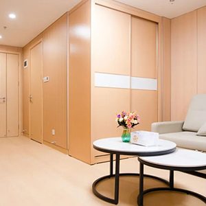
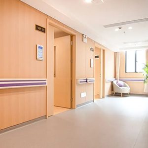
The International Medical Center and the International Center for Minimally Invasive Oncology Diagnosis and Treatment of Guangzhou Fosun Chancheng Hospital of Guangdong Pharmaceutical University (Guangzhou Xinshi Hospital) are located on the 5th floor of Inpatient Building 5, occupying approximately 1,500 square meters, and serve as the core area of the hospital's international medical services. The center offers an elegant environment and well-planned layout, with a reception area, specialist assessment rooms, dedicated treatment rooms, a dressing room, and multiple types of VIP wards.

Care pathway for international patients

1. Submit Materials

Send imaging studies and medical history via the form or WhatsApp

2. Specialist Assessment

The team reviews the condition and responds with feasible treatment options

3. Visit the Hospital

Assistance with travel arrangements, reception and admission procedures

4. Post-Treatment Follow-Up

Remote follow-up and health guidance after discharge

Frequently Asked Questions

What You May Want to Know First

Who is suitable for care at Oncology Ward 5?
The department mainly treats various types of advanced malignant solid tumors, including liver, lung, gastric, breast, colorectal and esophageal cancer, malignant lymphoma, malignant melanoma, tumors of the urinary system, gynecological system, head and neck, intracranial region, bone, soft tissue and retroperitoneum, and metastatic cancer, as well as certain benign tumors such as uterine fibroids. Suitability requires assessment by our specialists.
How does the multi-level drug-eluting microsphere technique differ from conventional intervention?
Conventional interventional technique requires selective catheterization to third-level branch arteries for local infusion and partial embolization; the programmed multi-level drug-eluting technique requires superselective catheterization to fourth-level arteries and their sub-branches, covering drug-eluting embolization from the peripheral vessels to the proximal main trunk. Specific suitability is determined by the physician.
How can international patients arrange treatment at the hospital?
You may first submit your medical records and imaging studies via the form or WhatsApp. After the team assesses your case, we will discuss feasible options with you. Once your visit is confirmed, we assist with travel, visa, accommodation and admission arrangements, and provide multilingual accompaniment services.
How long will I need to stay in hospital?
Length of stay varies depending on the type of disease, the treatment plan and individual recovery; it cannot be generalized. Details relevant to your individual case can be provided during the specialist assessment stage.
What does hyperthermia involve?
Non-invasive deep tumor hyperthermia uses an external radiofrequency hyperthermia unit, targeting above 39°C per session for approximately 40 minutes, with a recommended course of 6 sessions. The specific protocol is determined by the physician based on the patient's condition.

Submit Your Medical Records for a Preliminary Specialist Opinion

Please provide recent CT / MR imaging and pathology reports whenever possible — the more complete the material, the more targeted the assessment. We are committed to strictly protecting your personal information.

WhatsApp +852 4743 1548
💬WeChat: GaoCaoTangZY

Free Case Review

By submitting, you agree that we may use the above information to contact you; the information will be used solely for medical consultation purposes.

Important Notice

The content of this page is provided for medical reference and health education purposes only. It does not constitute any treatment recommendation, promise of outcome, or guarantee in the sense of medical advertising. The techniques, indications and case records presented here are intended solely to illustrate the clinical process and methods of the department. Any diagnosis and treatment must be determined by a licensed physician following an in-person consultation and evaluation; individual conditions differ and actual results vary from person to person — there is no therapeutic guarantee. Patient information in the case records has been anonymized and does not constitute testimonial or endorsement in the sense of advertising law. This page does not establish a doctor-patient relationship.

WhatsApp
💬